
26 Sep Cornea and Contact lens


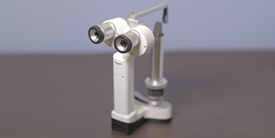
Slit lamp (Zeiss Ophthalmic, Germany)
Our institute is equipped with 2 different high resolution slit lamps by Zeiss and Appasamy.
allows doctor to examine front of the eye(Cornea,Anterior chamber,Iris and lens) under high magnification.
The use of specialised lenses(slit lamp bimicroscopy)makes it possible to see the back of the eye esp vitreous, retina and optic nerve in details and under very high magnification.
Imaging system(Camera attachment) helps in recording and comparing the detailed pictures of the eye during multiple visits of the eye, for objective assessment of improvements in the condition of the eye.


Corneal Topographer / Wave front Analyzer-Keratron Italy
Accurate computerized,in depth analysis of the front curvature and shape of the entire cornea.
Detects high order defects in the cornea, especially follwing corneal transplants or LASIK.
Helps in computerized fitting of contact lenses.
Allows detection of shape abnormalities of cornea like Keratoconus.
Essential for patients undergoing LASIK


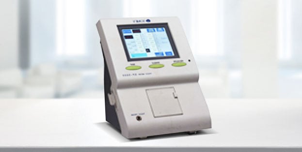
Corneal Pachymetry – (Tomey, Japan)
It Assesses thickness of the entire cornea. Ultrasound pachymetry is the gold standard in the measurement of cornea thickness since years.
Essential for patients undergoing LASIK, Corneal Collagen Crosslinking.
Heplful diagnostic tool in eyes with Glaucoma, to determine the target Intraoular pressure inside the eyes before and after treatment with antiglaucoma eyedrops.


Sorry, the comment form is closed at this time.